|
|
|
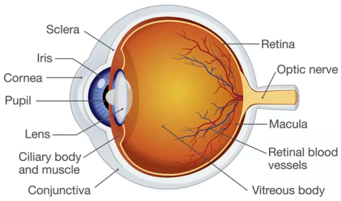
Vitreous gel, also called vitreous humor, is a thick, clear, gel-like fluid that fills the inside of the eye behind the lens and helps the eyeball maintain its shape. Two common eye conditions associated with the vitreous gel are floaters and flashes. Floaters are tiny clumps of debris or cells inside the vitreous moving in your field of vision. While these objects look like they are in front of your eye, they are actually floating inside. What you see are the shadows they cast on the retina. When the vitreous gel inside your eye rubs or pulls on the retina, you may see what closely resembles flashing lights or lightning streaks. Both floaters and flashes are usually harmless. However, if you notice any new or sudden increase in either symptom, you should consult your eye physician immediately because of the possibility of a torn retina.
|
|
|
|
The optic nerve, also called cranial nerve II, is a bundle of more than one million nerve fibers that carries visual messages from the retina to the brain. The optic nerve is associated with a number of diseases which typically causes permanent and potentially severe loss of vision. Examples of such diseases are glaucoma, optic neuropathy, viral infections, bacterial infections, and diabetes.
|
|
|
|
The macula is the small sensitive area in the center of the retina situated near the optic nerve that is responsible for central vision. The macula allows you to perform detailed tasks that require central vision such as reading or sewing. Damage to the macula, commonly known as macular degeneration, eventually results in central vision loss making reading or intricate work difficult or impossible without the use of special low vision optical aids.
|
|
|
|
Fovea Centralis, also known as fovia, is the center of the macular region of the retina that gives the sharpest central vision which is necessary for reading, watching television, driving, or any activity where visual detail is of primary importance.
|
|
|
|
The retina is the light-sensitive tissue lining the inner surface at the back of the eye. The retina converts light into electrical impulses that are sent to various visual centers of the brain through the optic nerve. A common problem related to the retina is retinal detachment. Retinal detachment occurs when the retina is pulled away from its normal position in the back of the eye allowing vitreous humor to leak under the thin tissue of the retina. When this occurs, vision is blurred and almost always causes some vision loss or blindness unless it is treated.
|
|
|
The iris is the colored part of your eye that serves as its first line of defense against ultra bright lights. Its primary function is to control the size of the pupil to regulate the amount of light entering the eye, ensuring the eye can see clearly in varying light levels. In bright light, the muscles within the iris constrict, causing the pupil to become smaller. This involuntary response reduces the amount of light that reaches the retina, protecting it from damage. In a dim or dark environment, these muscles dilate, causing the pupil to become larger. This allows more light to enter the eye, improving your vision in low-light conditions.
Eye color fun facts:
- Approximately 80% of the world’s population has brown eyes, making it the most common eye color globally.
- Green eyes are the rarest natural eye color, with only about 2% of the world’s population having them.
- People with light–colored eyes may be more light sensitive compared to those with dark–colored eyes. The iris pigment (melanin) helps block additional light in bright environments. With blue or green eyes, there is not a substantial amount of melanin to stop light from entering in the eyes. On the other hand, the dark pigment in brown eyes acts as natural sun protection in a way. Having brown eyes is by no means a substitute for wearing sunglasses, but it does help decrease the amount of harmful UV light that can lead to macular degeneration. For this reason, it is no surprise that people with blue eyes are more likely to develop this common eye condition.
-
Eye color genetics is far more complex than previously thought. Now outdated and oversimplified, the original theory suggested that eye color is controlled by a single gene with brown being dominant over blue. This led to the common belief that two blue–eyed parents could never have a brown–eyed child. However, that is not necessarily the case. With more than 50 genes from each parent influencing it, the child’s eye color has numerous possibilities including having a completely different eye color than either parent.
-
Interestingly enough, when children are first born, they usually have blue eyes because of the small amount of melanin the eye contains. However, melanin production generally increases during the first year of a baby’s life primarily due to accumulated exposure to light. The child’s permanent eye color is usually determined between 6 and 12 months; however, subtle shifts in eye color can continue for as long as three years as melanin granules continue to deposit and settle in the iris.
|
|
Ciliary body is a circular structure that is an extension of the iris. The ciliary body produces a water-like fluid in the eye called aqueous humor which protects the eye against harmful bacteria and transports vital nutrients to the eye. The ciliary body also contains the ciliary muscle, which changes the shape of the lens when your eyes focus on a near object.
|
|
|
|
The cornea is the clear front part of the eye that covers the iris, the pupil, and the anterior chamber. Because of its transparency, the cornea does not have blood vessels or a direct blood supply; therefore, it must exchange its nutrients and waste products through its front and back surfaces. The occurrences of corneal ulcers or keratitis are due to invasion by bacteria, fungi, or viruses causing infection and inflammation. This condition may cause severe pain and reduce visual clarity and are commonly treated with anti-bacterial or anti-fungal eye drops.
|
|
|
|
The pupil is the round opening located in the center of the iris of the eye. The muscular action of the iris adjusts the size of the pupil and controls the amount of light that can enter the eye. The round pupil appears black because most of the light entering the pupil is absorbed by the tissues inside the eye. Without any known cause, a small percent of the population has naturally unequal pupil size. However, causes of the size difference could include conditions such as glaucoma; head, neck, or eye trauma; or possibly an intracranial tumor that would require immediate medical attention.
|
|
|
|
The lens is a clear structure of the eye located behind the iris that helps focus light or an image on the retina. The lens, by changing shape, functions to change the focal distance of the eye so that it can focus on objects at various distances, thus allowing a sharp image of the object to be formed on the retina. Vision-threatening complications of the lens could include cataracts. A cataract is a clouding that develops in the lens of the eye, varying in degree from slight to complete obstruction of the passage of light. Cataracts typically progress slowly and are a natural process to aging.
|
|
|
|
The sclera is the tough, white outer coating of the eye that serves as the eye’s protection, also known as “the white of the eye”. Six tiny muscles connect to it around the eye and control the eyes movements.
|
|
|
Conjunctiva is a thin, transparent mucous membrane that lines the inside of the eyelids and covers the sclera, or better known as the white part of the eye. The conjunctiva does more than just provide immune defense, it has two other equally important roles to help keep the eye healthy. First of all, the conjunctiva protects the eye by producing a tear film that acts as a barrier against harmful bacteria, viruses, and dust and debris. Secondly, it keeps the eye well–lubricated, which ensures smooth eyelid movement and moisture retention. In turn, this promotes eye comfort by protecting it from dryness and irritation.
In a healthy eye, the conjunctiva is clear and colorless; however, when it becomes inflamed, either from an allergic reaction, a viral infection, or a bacterial infection, it can often lead to a red appearance in the eye. This very common condition known as conjunctivitis, or “pink eye”, often results in intense itchiness and swelling of the eyelids and a watery or pus-like discharge. Viral and bacterial conjunctivitis are contagious, while allergic conjunctivitis is not. Each type has different symptoms and requires different treatments.
|
|
|
|
Retinal blood vessels are the arteries, veins, and capillaries that work together to supply the retina with oxygen and nutrients; to carry deoxygenated, waste-filled blood away from the eye; and to remove waste products. The most serious medical conditions associated with the retinal vascular system include vein and artery blockages, diabetic retinopathy, and hypertensive retinopathy. These disorders, which can easily cause vision loss and even blindness, are often linked to other medical issues such as diabetes. Common risk factors include high blood pressure, high cholesterol, obesity, and smoking.
|